Vicor-CV SWIFT Medical Angiography X-ray System Features
1 High Definition and Stable Image
High resolution and wide-field images to support accurate and efficient diagnosis clinically
◆Digital flat panel detector
◆ Multiple panel models
◆ Adjustable, and wide-field
◆ Panel-digitized dynamic range up to 16 bit
◆ Accurate and clear details
◆ Resolution up to 1956×1956
2 Long-lasting and Efficient Performance
◆3.0M heat capacity X-Ray tube, having long-lasting and stable life.
◆ Liquid-metal lubricated bearing to improve heat dissipation efficiency.
◆ Triple focus spot, provide clear images for different clinical application.
3 Safe Radiation Protection
◆ Accurate working of the collimator to effectively reduce scattered rays
◆ Active radiation dose management and monitoring, and real-time and accurate recording of radiation dose and other information with RDSR during operation to realize effective supervision over the radiation dose
◆ Function of virtual collimator, to locate the checked site without X-ray egression
◆ Multiple dose models to meet different needs, and obtain high-quality images even with low dose
4 Professional Software Systems
Vicor CV Workstation
◆ 64-bit operating system, providing user-friendly interactive interface
◆ Accelerated real-time processing module with efficient GPU, and stable and reliable image
◆ Supporting multiple image processing tools such as automatic measurement, ventricular analysis and QCA analysis
◆ Real-time function of virtual beam limiter, helpful to reduce scattered rays and improve the safety of both physicians and patients
◆ Radiation dose report in compliance with IEC standard
◆ Safety design in compliance with network safety standard
Vicor AngioExpert
◆ Data transmission in compliance with DICOM3.0 and related standards
◆ Supporting data storage and migration management
◆ Efficient completion of vascular contour and morphological data analysis, and generation of QCA analysis report
◆ Supporting dynamic correction of stent position to enhance stent visualization
◆ Full-automatic seamless image splicing to expand diagnostic coverage and clearly display limb details
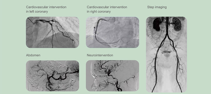
5 Clinical Application In All Departments

6 Stent Enhancement
As an image recognition technique, it is used to realize the motion compensation for the X-ray image to clearly and accurately display the fine structure of the stent, and help physicians immediately evaluate the release of the stent during the operation.
* This feature requires detailed consultation with sales
7 Swift 3D
With the perfect 3D reconstruction function, Vicor-CV SWIFT supports reconstruction techniques such as volume reconstruction (VR), multiplanar reconstruction (MPR), maximum intensity projection (MIP), and minimum intensity projection (MINIP). It has great advantages in analyzing the three-dimensional morphology and spatial relationship of blood vessels, and has great diagnostic value in interventional therapy for vascular system diseases, tumors, etc.:
1. It can avoid vascular structure overlapping, to make vascular images clearer and more accurate.
2. The three-dimensional relationship and tubal lumen structure between lesion sites and other normal tissues and organs can be observed at any angle.
3. The dosage of contrast agent, the time of diagnosis and treatment and surgery, and the exposure dose can be reduced.
4. Accurate measurement results can provide data for surgery and interventional treatment


 Email Us: