Matters Needing Attention, Detection Function and Process of Digital Subtraction Angiography
-
2022-10-25
-
LEPU

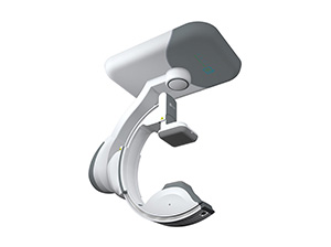
Digital Subtraction Angiography (DSA) is an angiography method with computer-assisted imaging, which is a brand-new X-ray examination technology that has been used in clinical practice since the 1970s.
Ⅰ. Precautions for digital subtraction angiography
1. The anesthesia method, contrast agent reaction, possible complications and postoperative care should be explained to patients and their families before surgery.
2. When intubating, a catheter with appropriate thickness and specifications, good smoothness and elasticity should be selected, and the operation should be gentle. The advancing and retreating of the guide wire and catheter should be carried out under fluoroscopic monitoring. It is not advisable to forcibly insert the guide wire in the case of resistance. The guide wire should not stay in the catheter for more than 90s. If the guide wire or catheter is found to be bent or broken, it should be corrected immediately, and papaverine can be given to relieve spasm in the case of vasospasm.
3. During the operation, attention should be paid to observe the patient's reaction, such as the reaction of speech, vision, hearing, and limb movements. If any abnormality is found, the operation should be stopped at any time. According to the situation, the general abnormal signs are mostly contrast agent reaction or vasospasm, which can be treated with antispasmodic drugs and antihistamine drugs after the angiography is confirmed.
4. 24 hours after operation, the patient's consciousness, limb activity and the pulse of the dorsal foot artery on the side of the puncture should be closely observed.
Ⅱ. The role of digital subtraction angiography
Commonly used in the diagnosis of cardiovascular and cerebrovascular diseases. It can not only clearly display the blood vessels of the internal carotid artery, vertebrobasilar artery, large intracranial blood vessels and cerebral hemisphere, but also measure the blood flow of the arteries, especially for aneurysm, arteriovenous malformation and other qualitative localization diagnosis, which is the best diagnostic method.
It can not only provide the exact location of the lesion, but also clearly understand the scope and severity of the lesion, providing a more reliable objective basis for surgery.
Ⅲ. Digital subtraction angiography examination process
1. Routine examination of blood, urine, stool, liver and kidney function, chest X-ray, electrocardiogram, and special inquiries about drug allergy, diabetes and asthma. Preoperative iodine allergy test and perineal skin preparation should be performed. When carotid artery cannulation or direct puncture of maxillofacial area is required, local skin preparation should be performed.
2. Anesthesia. Children need to be under the supervision of an anesthesiologist, and anesthesia such as ketamine can be applied. In adults, simple angiography can be performed under local anesthesia; if more complex embolization treatment is required at the same time, neuroleptic analgesia is required.
3. The Seldinger technique is used for intubation through the femoral artery. First choose the common carotid artery on the ipsilateral side, and then perform super-selective angiography of the external carotid artery branch. Angiography of the vertebral artery, the neck trunk or the contralateral neck is performed if necessary, depending on the lesion. Guidewire guidance can be used if the catheter cannot directly enter the vessel to be selected.
4. For those with negative iodine allergy test, meglumine can be used, and its concentration should not be too large, generally 40% to 60%. Non-ionic contrast agents should be used in patients with risk factors such as poor liver and kidney function, heart disease, diabetes, asthma, urticaria, eczema and other allergic diseases, as well as in children and the elderly.
5. After the angiography is completed, extubation, the femoral artery puncture point should be compressed for 10-15 minutes, and the pressure should be bandaged for 24 hours. The pressure should not be too heavy in children. The patient lay supine for 24 hours after surgery.
