What is an Embolic Coil?
-
2021-08-30
-
LEPU

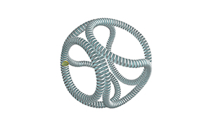
Coil embolization describes a minimally invasive surgical procedure to treat cerebral aneurysms and fistulas. Through a catheter inserted into the groin, a tiny coil is guided to the brain and separated from the catheter to prevent blood flow to the aneurysm or fistula. One or more platinum coils are left in place to prevent the aneurysm from rupturing. Reduced blood flow to the brain may affect binocular vision.
1. During operating the embolic coil, the femoral artery was touched at the place passing through the groin.
Neuroradiologists or neurosurgeons usually perform this operation in a hospital setting. The surgeon makes a small incision in the groin to access the femoral artery. The doctor uses dye to make the aneurysm visible in the computer image, and then passes the catheter through the artery. Once the aneurysm is close to the aneurysm, the surgeon releases the embolic coil from the catheter. The body creates blood clots around the coil, blocking blood flow.
An aneurysm represents a bulge or saccular artery wall in a weak patient. The protrusions can put pressure on the tissues and nerves in the brain, causing paralysis, or rupture, leading to stroke or death. Embolization coils can be used as a preventive measure after or before the aneurysm ruptures.
2. Neck pain when embolic coil is used may be a sign of aneurysm
Symptoms of aneurysm include headache, nausea or vomiting, upper back and neck pain. When these symptoms are present, doctors usually perform imaging tests to determine whether there is an aneurysm. When the patient cannot undergo brain surgery to prevent rupture, it is usually recommended to use detachable coils embolization.
An aneurysm represents a bulge or vesicle on the weak wall of an artery. The fistula defines the veins and arteries and reduces the delivery of oxygen-rich blood to the brain. These abnormal gaps usually cause eye pressure, which is a major symptom of glaucoma. Some fistulas can cause diplopia, pain, and abnormal sounds in the ears, such as buzzing.
If an aneurysm mass is large or appears at the base of the skull, balloon occlusion may play a role. This is another option when embolization coils cannot be performed due to the size or location of the aneurysm. This operation uses an inflated balloon to restrict blood flow. The procedure is similar to the detachable coil embolization of the femoral artery cannula. The risk of this operation is considered low, but a stroke may occur during the embolization of the coil, the patient's legs or arms may be weak, and language and vision problems may also occur.
3. The embolic coil for treatment of cerebral aneurysms
Neurosurgeons can use brain imaging to locate fistulas and aneurysms in the brain. After placement, the patient remains lying flat for 8 hours or more to allow the femoral artery to heal. They usually go home after a day or two. A few months later, angiography may be performed to determine whether the embolization coil is still in place. Neurosurgeons usually perform detachable coil embolization to treat cerebral aneurysms.
